Saturday, 17 April 2021

Lupine Publishers| Risk of Antimicrobial Resistance Development from Pet Animals to Humans: Case of Enterobacteriaceae Family

 Lupine Publishers | LOJ Pharmacology & Clinical Research


Antimicrobials are vital instruments for the treatment of contagious bacteriological infections in pet animals, as well as in humans. The demise of the effectiveness of antimicrobial substances can honestly deal with pet animal health and human health. A necessity for the enhancement of innovative antimicrobials for the treatment of multiresistant infections, specifically those caused by Gram-negative bacteria, has been recognized in human medicine, and an imminent subsequent demand in veterinary medicine is required. A distinctive feature associated with antimicrobial resistance and the risk of resistance development in pet animals is their close interaction in conjunction with humans. This generates chances for interspecies transmission of resistant bacteria. This review aims to recapitulate the current information on the use and indications for the Enterobacteriaceae family in pet animals and the spread of antimicrobial resistance among pet animals and their owners. The critical antimicrobial resistance microbiological threats from pet animals that directly or indirectly may cause adverse health effects in humans are carbapenemase-producing Enterobacteriaceae bacteria such as Escherichia coli, Klebsiella spp., Enterobacter spp. and Salmonella spp.

Keywords: Antimicrobial resistance; Antibiotics; Public health; Microbiology

Introduction

Throughout the last half of the century, the quantity of pet animals in contemporary civilization has considerably augmented, and a modification in their public part has arisen [1]. Awareness of their welfare has grown because of the close interaction between pets and their owners [2]. Humans could develop antimicrobialresistant bacteria or the consequent resistance genes from food animals [3,4] and interaction with their pet animals [5,6]. Enterobacteriaceae and multidrug-resistant Gram-negative bacteria have become apparent in healthy and sick pets, suggesting a possible threat of the spread of these bacteria to humans from contaminated or inhabited pet animals [7]. Additionally, there is also the possibility to transfer resistance genes vice versa. To evaluate the hazards contained by the framework of treatments for new antimicrobials for pet animals, could arise a necessity for further data requirements concerning antimicrobial resistance [8]. Antimicrobials are used commonly in the routine procedure for medicinal and preventative reasons in pet animals [9]. Nevertheless, antimicrobial intake data for dogs and cats are repeatedly deficient and typically addressed to drug company trades [10]. Even if trade data provide a challenging assessment of antimicrobial consumption’s enormity, data on the utilization of antimicrobials in various species are deficient [11]. Pet animal sales of antimicrobials are a small percentage of the global sales example of animals’ antimicrobial agents.

Various antimicrobial products approved for human use are also used in pet animals in the treatment of the “cascade” [12]. Prevalent use of broad-spectrum antimicrobials has been described in pet animal practice in Europe (Figure 1). The most used antimicrobials for dogs and cats are β-lactams, for example, amoxicillin and amoxicillin combined with clavulanic acid [13]. Shortage of verified diagnosis might take the lead to the abuse of antimicrobials [14]. Antimicrobial management has occurred to treat disorders in which effectiveness has not been demonstrated, for instance diarrhea in dogs for which antimicrobial treatment is generally not proposed [15,16]. Multidrug resistance bacteria have been described in pet animals, every so often cruelly conceding the therapy result. Since restricted reconnaissance and understanding of the zoonotic transmission of antimicrobial resistance between humans and pet animals, the level of spread and significance for public health is inadequately appreciated [17]. Within the next, the critical drug-resistant bacteria are assessed and the indication for their transmission among humans and their pet animals.


Enterobacteriaceae as a Significant Public Health Concern in Human Medicine

Go to

Representatives of the Enterobacteriaceae family comprise numerous species, for instance, Escherichia coli, Enterobacter spp., Salmonella spp., and Klebsiella spp. [18]. Countless bacteria belonging to these species are symbiotic organisms of the digestive tract [19]. Increasing antimicrobial resistance among Enterobacteriaceae is evolving as an essential public health apprehension in human medicine. Enterobacteriaceae, which produce ESBLs, extended-spectrum cephalosporinases, and plasmid-mediated AmpC β-lactamases (ESBLs) are particularly important. Around are quite a few articles on ESBL-producing bacteria in pet animals [6]. Escherichia coli, Salmonella spp., Enterobacter spp., and Klebsiella spp. as Potential Hazards Over the years, E. coli was reported continuously from the first recorded case in Japan in pet animals, followed by the emergence in humans in following years, to the incidence in ESBL-producing uropathogenic E. coli from pet animals in Spain [20]. Since that time, the number of statements relating to E. coli ESBLs in pet animals has grown precipitously [21]. CTX-M enzymes have developed a swiftly expanding family of ESBLs in bacteria from human infections. In pet animals, equally clinical and commensal isolates of E. coli frequently generate CTX-M type β-lactamases [22]. E. coli has lately become known as a global pandemic replicate in humans [23]. Descriptions of clinical infections in animals caused by E. coli are merger, which may be since its detection requires genotypic techniques. Various clinical E. coli isolates from pet animals are like human clinical E. coli isolates founded on their virulence genotype, and resistance characteristics, etc. Many E. coli strains such as ST156, ST405, ST410, and ST648 could be found both in humans and their pets [24]. The detection of duplicates in humans and dogs and cats may suggest their transmission through direct contact. Such transmission could likewise be a related component to the prompt and effective spreading of E. coli, even though, between humans, the incredibly critical transmission path is almost certainly "hand to hand" [25]. Salmonella spp. have been correlated with epidemics of nosocomial intestinal infections in pet animals in veterinary clinics and an animal sanctuary-some of the outburst as well engaged veterinary organization and other people in connection with pets [26].

In cases like this, pet animal sanctuaries could work as foci of transmission for Salmonella spp. among humans and animals if acceptable control measures are not provided [27]. Information on antibiotic resistance phenotypes and genotypes of Salmonella spp. in animals and humans in different countries and geographic regions is necessary to combat the spread of resistance [28]. This will improve the understanding of antibiotic resistance epidemiology, tracing new emerging pathogens, assisting in disease treatment, and enhancing the prudent use of antibiotics. However, the extent of antibiotic resistance in foodborne pathogens and humans in many developing countries remains unknown [29]. Among 25526 recorded isolates of salmonellae, 5086 isolated from humans, and 20440 from animals in 1994 and 1997 in France, the antibiotic resistance phenotype was determined for all human and 5336 animal isolates. In Salmonella enterica serovar typhimurium, one of the two most frequently isolated serovars from humans as well as animals, resistance to ampicillin was observed in 61% of both human and animal isolates in 1994 and in 73% of human and 53% of animal isolates in 1997.

During these periods, resistance to co-amoxiclav was between 45% and 66% for both types of the isolate. Resistance to ampicillin was associated with resistance to streptomycin, spectinomycin, sulphonamide, tetracycline and chloramphenicol in over 70% of isolates [30]. Resistance to ampicillin as well as co-amoxiclav never exceeded 7% in Salmonella enteritidis. While Salmonella hadar was practically absent among the human isolates in 1994, this serovar was the third most frequent in 1997, and at that time, 92% were resistant to nalidixic acid. Among the animal S. hadar isolates, the prevalence of resistance to nalidixic acid increased from 3% in 1994 to 72% in 1997. None of these isolates manifested high-level resistance to ofloxacin. The levels of resistance to aminoglycosides (<3%) and trimethoprim-suphamethoxazole (<14%) remained practically unchanged in all three serovars. The resistance markers of 463 ampicillin-resistant S. typhimurium isolated in 1997 were determined. Among the 24 phenotypes observed, six multiresistant phenotypes, representing 82% of these isolates (as compared with 80% in 1994), were associated with the PSE-1 gene typically found in the lysotype DT104 of this serovar [30]. Being as by E. coli, extended-spectrum cephalosporinases and plasmid-mediated AmpC β-lactamases producing strains of Salmonella spp. are of disquiet [24]. Antibiotic of the cephalosporin type licensed for use in veterinary medicine resistance was detected in more than 10% of cats and 21% of dog Salmonella spp. isolates with detected β-lactamases, respectively [31]. The main issue and problem are that there is limited knowledge of ESBLs in other Enterobacteriaceae of pet animals. Klebsiella spp. from the human epidemic clone was isolated from dogs and cats in Spain [32]. It was found to be highly resistant to aminoglycosides due to the ArmA methyltransferase. The emergence and clonal spread of Klebsiella spp. in dogs were first reported in Germany [33]. While in Singapore, an analysis of 186 diagnostic reports collected from a veterinary clinic between 2014 to 2016 showed that sick companion animals could carry bacteria of significance to human health [34]. Among the 186 specimens submitted, 82 showed polymicrobial growth (45%, 82/186), and in total, 359 bacteria were isolated. Of the 359 bacteria reported, 45% (162/359) were multi-drug resistant, and 18% (66/359) were extended-spectrum-β-lactamase species. Resistance to broad-spectrum antibiotics was also observed among individual species. Namely, methicillin-resistance among Staphylococcus pseudintermedius (63%, 32/51) and Staphylococcus aureus (50%, 4/8); fluoroquinolone-resistance among Escherichia coli (40%,17/42) and carbapenem-resistance among Klebsiella pneumoniae (7%, 2/30) were noted [34].

This analysis suggests that sick pets may contribute to the pool of clinically relevant antibiotic-resistant bacteria and play a role in the spread of antibiotic resistance. Antibiotic-resistant bacteria such as Klebsiella pneumoniae are common in the digestive tract and upper respiratory tract of animals and humans [35]. Several studies have shown that this bacterium is found in humans and in animals, one of which is pigs that are known to be a reservoir for the spread of this bacteria [36]. Not only in pigs, but this antibiotic-resistant bacterium is also known to be found in other food-producing animals, as well as in pet animals. Many cases of Klebsiella pneumoniae in humans have been reported, but Klebsiella pneumoniae in humans related to animals or strains related to animals and humans were also reported [37]. Control and prevention are needed to prevent the spread of antibiotic-resistant bacteria from animal to animal, animal to human and vice versa, and the surrounding environment.

Conclusion

In humans, the control of resistance is based on hygienic measures: prevention of cross-contamination and decreased antibiotic usage. In animals kept together, sanitary measures, such as prevention of oral-fecal contact, are hardly achievable. Consequently, lessening the need for antibiotics is the only possible way of managing resistance in pet animals. This can be achieved by improving pet animal welfare systems and eradicating or vaccinating against infectious diseases. Furthermore, eliminating antibiotics as preventive measures in pet animals would decrease antibiotic use and minimize transmission from animal to human resistance. This would not only diminish the public health risk of dissemination of resistant bacteria or resistant genes from pets to humans but would also be of significant importance in maintaining the efficacy of antibiotics in human medicine and veterinary medicine as well. A more extensive study to better understand the extent of distribution and the factors affecting antibiotic-resistant bacteria’s transmission to and from pets is more than necessary.

Acknowledgment

This research was supported by COST Action “European Network for Optimization of Veterinary Antimicrobial Treatment”, grant number CA18217.

https://lupinepublishers.com/pharmacology-clinical-research-journal/fulltext/risk-of-antimicrobial-resistance-development-from-pet-animals-to-humans-case-of-enterobacteriaceae-family.ID.000138.php

https://lupinepublishers.com/pharmacology-clinical-research-journal/pdf/LOJPCR.MS.ID.000138.pdf

For more Lupine Publishers Open Access Journals Please visit our website: https://lupinepublishersgroup.com/
For more Pharmacology & Clinical Research Please Click
Here: https://lupinepublishers.com/pharmacology-clinical-research-journal/
To Know more Open Access Publishers Click on Lupine Publishers

 
Follow on Linkedin : https://www.linkedin.com/company/lupinepublishers
Follow on Twitter   :  https://twitter.com/lupine_online

Friday, 9 April 2021

Lupine Publishers | Advances in Chemical Composition and Biological Activity of Mexican Propolis

 Lupine Publishers | LOJ Pharmacology & Clinical Research


Abstract

Propolis is a product created by honeybees from vegetal sources. Nowadays it is considered a medicinally or functional food because of the phytochemicals contained in it. Numerous papers have been published reporting the chemical composition of different countries around the globe due to its highly variability(phenolic compounds, sugars, terpenoids). Due to the variability in its chemical composition, various biological activities have been reported, among them are anti-inflammatory, antimicrobial, anticarcinogenic, among other. In Mexico has history on beekeeping that was modified by the Spanish conquest but endures to these days. Therefore, it is important to exploit the characteristics of the propolis of our region in order to know both their composition and beneficial properties.

Keywords: Propolis; Honeybees; Flavonoids

Introduction

Propolis is resinous material produced by honeybees, which collect secretions from plants organs and then they are mixed with beeswax and salivary enzymes [1]. Propolis color may vary from light yellow to dark brown [2]. Bees produce propolis to fill cracks in the hive, to regulate the temperature and embalm invaders [3]. The general composition comprises resins (50%), waxes (30%), essential oils (10%) pollen (5%) and other organic compounds (5%) [4]. Some compounds reported in the resin are phenolics, esters, flavonoids, aldehydes and alcohols. This natural product has attracted the attention of researchers because of its biological activity and the diversity of its composition. Since 1970’s, scientists around the world have reported the chemical composition and biological composition of propolis from different countries such as Czeck Republic, Bulgaria, Croatia, Portugal, China, Netherlands, Italy, France and Brazil, among others. However, in Mexico little has been explored of the propolis of this region despite that apiculture has been an ancient activity. Pre-Colombian cultures raised stingless bees for food, medicinal and religious purposes. In the tropical region of Mexico, Mayans bred honeybees such as Melipona beechii, Melipona domestica, Melipona fulvipes and Trigona sp, in order to collect honey and wax [5]. Spanish conquerors brought with them the European bee, Apis mellifera, who partially replaced the native Mexican bees in recollection of bee products. Nowadays, Apis mellifera is the main honeybee exploited in Mexico for honey, beeswax, pollen, royal jelly and propolis. Propolis is considered only a side product derived from the hive, therefore there is no reliable source to measure the production of Mexican propolis [6]. However, it is known that the Peninsula of Yucatan has been the most productive from 2000 to 2012, in terms of honey production [7]. Propolis is commercialized in Mexico in regional markets and local producers in its raw form, but mainly it is combined with honey to make hard candies or hydroalcoholic solutions (Figure 1). The quality of propolis or propolis products is dictated by Mexican regulation NOM-003-SAG/GAN-2017 “Propóleos, producción y especificaciones para su procesamiento”. This normative comprises a qualitative test of flavonoids and phenolics, oxidation index, quantification of phenolics, quantification of flavonoids, antioxidant activity (DPPH bleaching assay) and antimicrobial activity on three bacteria. It is worthy to mention that before the NOM-003-SAG/GAN-2017, there was no regulation to ensure the quality of propolis.

Chemical Composition

Research on propolis has revealed over 500 constituents: phenolic compounds, fatty acids, sugars, minerals and terpenoids [8-11]. It is known that propolis composition varies greatly depending on the geographical origin, the botanical source, the time and method of harvest and solvent used in extraction [12-13]. The Ministry of Agriculture and Rural Development has divided Mexico into five beekeeping areas, in accordance to the honey production and geographical distribution: Altiplano, Pacific Coast, Gulf, North and Peninsula of Yucatan [14]. We can assume that propolis composition would have a pattern for each region as honey does. To date, there are reports of composition from the North (A, B, C, D, E, F and G), Altipano (H), Gulf (I), Pacific Coast (J), and Peninsula of Yucatan (K, L, M and N) (Figure 2).


Phenolics and Phenolic Esters

Phenolics are natural constituents of plants that are characterized by the presence of hydroxy substituents in their structures. Together with total flavonoid content, phenolics content in propolis is another quality characteristic in Mexican regulation NOM-003-SAG/GAN-2017, which minimum content must be 5%w/w. Only samples from Caborca, Ures, Pueblo de Alamos, Aquiles Serdan, Parral, Gomez Palacio, Fresnillo, Silao and Solidaridad were studied for phenolic content, from which the Solidaridad sample did not fulfilled the Mexican regulation. Sample from Solidaridad showed no content of phenolics since the major compounds were terpenoids. Argentinean and French propolis possess similar phenolic content [15-16].

Flavonoids

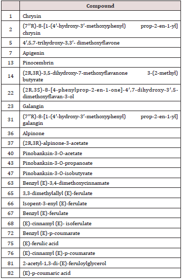
Table 1: Flavonoids and phenolics isolated from Sonora propolis and tested for antiproliferative activity.

lupinepublishers-openaccess-journal-pharmacology-clinical-research-journal

Flavonoids are the most reported compounds in propolis around the world. The content of flavonoids is considered to reflect the quality of the propolis, as the Mexican regulation NOM- 003-SAG/GAN-2017 stablishes the requirement of minimum of 0.5%w/w of flavonoids. However, only samples collected in the regions Aquiles Serdan, Parral, Gomez Palacio, Fresnillo and Silao were assessed for this parameter and found to fulfill the content of flavonoids. Table 1 depicts the types of flavonoids isolated from the propolis samples. Pinocembrin was the most reported compound in Mexican propolis, in ten out of fourteen samples, followed by Chrysin in six out of fourteen samples. Both flavonoids, together with galangin, kaempferol, rhamnetin and epoxypinocembrin chalcone, have been identified in poplar bud exudates [17]. In fact, pinocembrin is considered the marker of poplar propolis [18], therefore it is found in propolis from Spain, France, China, Portugal, Rumania, Croatia, Turkey, New Zealand, Poland, Argentina [12,19- 27].

Pterocarpans

The major components of South American red propolis (eg. Cuba and Brazil) are isoflavans, isoflavons and pterocarpans [28]. The sample of the region Champoton turned to be the only one containing pterocarpans and isoflavonoids, hence it could be excluded from the classification of poplar type and considered as red propolis.

Triterpenoids

Triterpenoids are natural occurring compounds in plants, which have anti-inflammatory, hepatoprotective, analgesic, immunomodulatory, antimicrobial, antimycotic, virostatic, and tonic effects [29]. Hunucma and Solidaridad samples contained exclusively triterpenes, this fact excludes them from the poplar type, red type or any other, since there is no type of propolis defined solely on triterpenes. However, the high content of triterpenes has been found in propolis from Ethiopia, being α- and β-amyrins and amyryl acetates the major compounds [30]. It is also similar to geopropolis from Mani (See Section Geopropolis). Botanical origin Depending on the ecosystem, honeybees will collect resins from different source plants, choosing appropriate representatives of the local flora [31].

Salatino and coworkers [18] made a classification of propolis, according to its geographic zone, chemical composition and botanical source reported in literature, into five types: I, from Populus section Aigeiros; II, from Baccharis dracunculifolia; III, from Clusia; IV, from Macaranga and V, Cupressaceae. According to this classification, Mexican propolis belongs to type I propolis, the temperate poplar propolis. Bees seem to gather the resins from Populus species and their hybrids. As for the tropical regions, the plant sources are rich of prenylated benzophenones, diterpenes and flavonoids [4]. As mentioned before, Mexican propolis falls into the classification of poplar type or type I. However, this is true for samples North, Pacific Coast and Gulf, since Pinocembrin and other flavonoids and phenolics were found as major constituents. The most common source from these compounds is the Populus genus, in fact the composition of Populus bud exudates have been studied to corroborate the botanical origin [17]. In Mexico is not well reported the species and abundance of Populus present throughout the country. Nevertheless, it is known that flavonoids such as pinocembrin, pinobanksin 3-acetate, tectochrysin, galangin, and chrysin have been found in Populus nigra [28]. Secondary sources visited by Apis mellifera are birch, alder, oak, pine and hazel. The southeast region of Mexico has tropical weather which is directly reflected on the chemical composition of propolis. Champoton, Hunucma, Mani and Solidaridad samples have a different composition from the rest of the country (See Section Chemical Composition), since these regions are considered to have a tropical weather. In tropical or subtropical areas, poplar trees are scarce, as a result, bees visit another type of trees. Two potential sources used by the bees for their production of propolis in Yucatan and Quintana Roo are Bursera simaruba and Lysiloma latisiliquum [20,32]. Dalbergia is another probable source of resin material in this region, because some compounds isolated from these samples (eg. (-)-Mucronulatol, (+)-Vestitol and (-)-Melilotocarpan A) are related to Dalbergia exudates and Caribbean and Brazilian propolis.


Biological Activity

Poplar type propolis is characterized by flavonoids, phenolic acids and their esters as bioactive constituents [28]. Particularly, flavonoids are associated to a broad spectrum of health effects because if their antioxidant, anti-inflammatory, anti-mutagenic, antimicrobial, anticarcinogenic and vascular activities [33].


Anticancer

Silao extract showed an inhibition of proliferation of rat C6 glioma cell line comparable to temozolamide, on the other hand, it did not efficiently inhibit human cervical cancer cell lines (HeLa, SiHa, and CaSki) proliferation compared to cisplatin [34]. Alday and coworkers [35] tested the antiproliferative activity of the methanolic extract of Sonora and the flavonoids isolated from it, on B cell lymphoma cell line M12.C3.F6. The methanolic extract exhibited an inhibitory concentration (IC50) of 20.6±0.5μg/mL, while the IC50 of the flavonoids pinobanksin-3-O-propanoate, pinobanksin- 3-O-butyrate, pinobanksin-3-O-pentanoate, pinobanksin, CAPE, galangin, chrysin ranged from 17.3μM to 76.6μM. Moreover, the extract showed to induce apoptosis through loss of mitochondrial membrane potential and activation of caspase 3, 8 and 9. Flavonoids and phenolics listed in Table 1 were isolated. These compounds were evaluated for the preferential cytotoxicity on PANC-1 human pancreatic cancer cells, cytotoxicity was expressed as PC50 [36]. The PC50 ranged from 4.6 to 98.9μM, showing a great potential for antiproliferative activity.

Hernandez and coworkers [37] tested the antiproliferative effect of Caborca, Ures and Pueblo de Alamos samples on human lung carcinoma A-549, human colonic adenocarcinoma LS 180, human cervix carcinoma HeLa , normal subcutaneous connective tissue NCTC clone L 929, murine B-cell lymphoma M12.C3.F6 and macrophage, Abelson murine leukemia virus transformed RAW. The components of propolis chrysin, xanthomicrol, acacetin, pinocembrin, naringenin, hesperetin, rutin, pinobanksin-3-acetate and CAPE, were tested as well. Pueblo de Alamos extract showed the best antiproliferative activity in all the cell lines tested in a range of 0.8 to 53.6 (μg/mL). Only CAPE, galangin, chrysin and xanthomicrol showed antiproliferative activity on most of the line cells (IC50 3.2- 95.4 μM), mainly on L-929, M12.C3.F6, RAW and, HeLa. These flavonoids could be partially responsible for the antiproliferative activity of propolis extracts. Flavonoids can exert anticancer effect through various mechanisms such as inactivation of oxygen radicals (antioxidative), binding to electrophiles, induction of phase 2 detoxification enzymes, increase in apoptosis, inhibition of cell proliferation and inhibition of lipid peroxidation [38].


Anti-Inflammatory

Chiapas (J) extract showed anti-inflammatory activity comparable to indomethacin (IC50 1.21 and 0.84 mg/ear, respectively) in the model of ear edema in mice by 12-O-tetradecanoylphorbol- 13-acetate. Besides diminishing histologic signs of inflammation, J decreased the activity of myeloperoxidase [39]. The flavonoids pinostrobin, izalpinin, cinnamic acid, kaempferol, 3,3-dimethylallyl caffeate, isopent-3-enyl caffeate, 3,4-dimethoxycinnamic acid and rhamnetin did not show antiinflammatory activity. Whereas pinocembrin had comparable effect to celecoxib (IC50 2.53μmol/ear and IC50 0.91μmol/ear, respectively) and to indomethacin (91.09±3.66% inhibition and 91.09.09±0.006%, respectively). The authors attribute to pinocembrin the anti-inflammatory effect observed in the propolis extract because of its high myeloperoxidase inhibition. Research on pinocembrin has revealed that it downregulates TNF-α, IL-1β, and IL-6, also suppresses IκBα, JNK and, p38MAPK [40]; which are some mechanisms that additionally explain the anti-inflammatory effect.


Antidiabetic

Chihuahua (D) propolis extract administered to streptozotocin induced diabetic mice reduces blood glucose levels and increases serum insulin levels. The pancreatic islets were found to contain insulin in contrast to the diabetic controls. There was found that the activity of antioxidant enzymes superoxide dismutase, catalase and glutathione peroxidase were restored in the treated mice in compare to non-treated mice [41]. The observed hypoglycemic activity could be the result of the secretagogue and antioxidant effects of the extract. Pinocembrin has a potential therapeutic use in diabetic nephropathy, a chronic complication of diabetes mellitus. When pinocembrin was administered to streptozotocin-induced hyperglycemic rats, before diabetic nephropathy establishment, it was able to improve survival and, to partially decrease blood glucose, lipids and renal function. These results correlated with a decrease in oxidative stress. However, when pinocembrin was administered once renal structural damage was stablished, pinocembrin improved triglycerides and LDL, but it worsens all renal function parameters due to increases ROS [42].

Iranian propolis has similar effects to Mexican propolis, on streptozotocin-induced hyperglycemic rats. The ethanolic extract of Iranian propolis avoided body weight loss and, high FBG and MDA content, as well as reducing GBM thickness and glomerular area. These results indicate that propolis protection was due to its antioxidants properties [43]. Various flavonoids have been studied for its antidiabetic properties. Naringenin has showed to inhibit of intestinal α-glucosidase, to improve insulin sensitivity and glucose tolerance [44-45]. On the other hand, chrysin inhibits TNF-α pathway and downregulates the expression of TGF-β, fibronectin and collagen-IV proteins [46]. These flavonoids, reported to be present in Mexican propolis, with their aforementioned mechanisms and their known antioxidative activity, could explain the antidiabetic effect described for the Chihuahua propolis.


Antimicrobial

Ethanolic extract of Silao (H) propolis and, the isolated flavonoids and phenolic acids were tested against oral pathogens Streptococcus mutans, Streptococcus oralis, Streptococcus sanguinis, and Phorphyromonas gingivalis. The inhibitory effect on the microorganisms ranged between 125-500μg/mL for the propolis extract and 128-1024μg/mL for the isolated compounds. Chiapas propolis (J) showed moderate inhibitory effect (MIC=250μg/mL) on Mycobacterium tuberculosis H37Rv in the Resazurin Microtiter Assay [39]. Sonoran propolis (Caborca, Ures and Pueblo de Alamos) samples were investigated for the effect on clinical isolates of Staphylococcus aureus. Ures sample had the highest inhibitory activity (MIC 200μg/mL) while Pueblo de Alamos showed no activity [47]. These samples also inhibited the growth of various Vibrio species such as cholerae (serotypes Inaba, non-O1, Ogawa), vulnificus, alginolyticus, fluvialis and parahemolyticus, in a range of 50-200μg/mL. Furthermore, propolis constituents were tested against V. cholerae, showing high (galangin and CAPE), moderate (pinocembrin, hesperetin and naringenin) and low (chrysin and acacetin) activity [48].

Quintero-Mora and coworkers [49] investigated the inhibitory activity on Candida albicans of various propolis from State of Mexico, Veracruz and Puebla not shown in Figure 2, fresh prepared and commercial extracts. The MIC was highly variable and ranged from 0.6 to >10 mg/mL. Recently Al-Ani et al. [50] tested propolis collected in Germany, Ireland and Czech Republic on Grampositive (Staphylococcus aureus, Staphylococcus saprophyticus, Staphylococcus epidermidis, MRSA/NCTC, VRE Van B, Streptococcus pyogenes, Streptococcus pneumoniae, Streptococcus oralis, Streptococcus agalactia, Streptococcus thermophilus, Bacillus subtilis, Enterococcus casseliflavus), Gram-negative bacteria (Klebsiella pneumoniae, Klebsiella oxytoca, Escherichia coli, Escherichia coli O157:H7, Pseudomonas aeruginosa, Salmonella choleraesuis, Shigella flexneri, Haemophilus influenzae, Acinetobacter baumannii, Burkholderia cepacia, Enterobacter clacae, Yersinia enterocolitis) and fungi (Candida albicans, Candida glabrata, Candida parapsilosis, Candida tropicalis, Candida krusei). The MICs for Gram-positive bacteria ranged from 0.08mg/mL to 5mg/mL; 0.6mg/mL to 5mg/ mL for Gram-negative and, 0.1mg/mL-5mg/mL for fungi. Thus, the European propolis showed higher inhibitory effect than that observed in Mexican propolis. Antimicrobial activity of propolis is attributed to the phenolic content and particularly, to the flavonoids galangin and pinocembrin [51]. The reported mechanisms are membrane damage, inhibition of acid nucleic synthesis, and inhibition of energy metabolism [52].


Antiparasitic

Sonoran propolis (Caborca, Ures and Pueblo de Alamos) samples were investigated for the effect on Giardia lamblia trophozoites. Ures sample showed the highest activity (IC50=63.8μg/mL), Pueblo de Alamos had low activity (IC50=222μg/mL) and Caborca did not show significant inhibition on the trophozoite growth. In the microscopic analysis of the trophozoites, there were morphological changes on the trophozoites treated with B compared to the control trophozoites, as well as cellular debris. The six main constituents of Sonoran propolis (CAPE, naringenin, hesperetin, pinocembrin, chrysin and, rutin) were tested on G. lamblia trophozoites, being CAPE the most active (IC50 63.1±0.9 μg/mL) and, hesperetin (IC50 149.6±24.8μg/mL) and pinocembrin (IC50 174.4±26.9μg/mL) the weakest ones [53]. European propolis extracts collected in different regions of the United Kingdom, Bulgaria and Lithuania, showed activity against human and bee parasites. The extracts decreased the survival of wild-type and resistant strains of Leishmania mexicana with a EC50 0.1-5.67μg/mL and 0.29-1.55μg/mL, respectively; wild-type and resistant strains of Trypanosoma brucei with a EC50 3.67-18.4μg/mL and 2.5-25.0μg/mL, respectively; Trypanosoma congolense with a EC50 1.96-35.7μg/mL and, Crithidia fasciculata with a EC50 2.58-23.8 μg/mL. Orthogonal partial least squares analysis of the propolis activity and the LC-MS profile, revealed that butyrate ester of pinobanksin is related to the high activity against Trypanosoma brucei, methyl esters of chrysin are related to high activity against Trypanosoma congolense, methyl esters of galangin and pinobanksin are related against Crithidia fasciculata, but no compounds were found to be related to the anti-Leishmanial activity [54]. The activity of Mexican propolis and European propolis may not be comparable since they inhibit different parasites, however, they both bring out the potential of temperate propolis and flavonoids against parasites.


Geopropolis

Stingless bees mix resinous material from plants, bees wax and soil to form geopropolis. This type of bees, also called Meliponinos, is present in all tropical zones over the world and around 400 to 500 species are known [55]. In Mexico 46 species have been identified, being Plebeia, Trigona, Melipona and Trigonisca the most diverse genera [56]. Stingless beekeeping was present in the cosmogony and mythology of Mesoamerican civilizations, as well as a source of staple food [57]. The most exploited species of Meliponinos, for their honey and wax, are Melipona beecheii in the Mayan area and Scaptotrigona mexicana in the Nahua area [56]. Despite the important historical background of stingless bees in Mexico, there is scant research on the propolis made by Meliponinos native to this country. The available research on geopropolis is from Brazil yet is not as abundant as the propolis research. Geopropolis differs from propolis because of the mineral and soil content, and the absence of plant trichomes of the former, despite of this, geopropolis has similar functions in the hive [58]. Biological activities reported for geopropolis are gastroprotective, antimicrobial, antioxidant, anticancer, anti-inflammatory, sedative, antimutagenic [59-63].

Regarding the chemical composition, polyphenols, phenolic acids, flavonoids, hydrolysable tannins, triterpenoids and, saponins [60,64-65]. Pentacyclic triterpenes were the main compounds identified in Melipona beecheii geopropolis from Mani (L), for instance lupenone, α-amyrin and β-amyrin [66]. Volatile compounds comprise approximately 10% of the propolis constituents and they confer odor and pharmacological effects. Pino et al. [67] compared the volatile fraction of propolis from Apis mellifera and Melipona beecheii from Yucatan, they were able to establish characteristic compounds of each type of propolis. Honeybees propolis’ exclusive volatiles were styrene, phenylacetaldehyde, trans-sabinene hydrate, nonanal, decanal, 2-undecanone, cyperene, cis-α-bergamotene, massoia lactone, ar-curcumene, cis-calamenene, cadina-1,4- diene, α-cadinene, β-eudesmol, α-bisabolol, neryl linalool, geranyl linalool, manoyl oxide, kaur-16-ene, pentacosane and heptacosane. Stingless bees geopropolis’ exclusive volatiles were santolinatriene, 6-methyl-5-hepten-2-one, α-phellandrene, δ-3-carene, p-menth- 1-ene, p-cymenene, α-pinene oxide, chrysanthenone, trans-pmentha- 2,8-dien-1-ol, cis-limonene oxide, verbenyl ethyl ether, germacrene A, α-dehydro-ar-himachalene, β-vetivenene and humulene epoxide I. However, both samples had in common the compounds α-pinene, β-pinene, trans-verbenol, α-copaene, β-bourbonene, β-caryophyllene, spathulenol and caryophyllene oxide. Geopropolis from Coatepec, Veracruz was subjected to HS-SPME-GC-TOF-MS analysis to identify volatile components. The major compounds identified in the total current ion were β-fenchene, styrene, benzaldehyde, (Z)-ocimenone, α-pinene, m-cymene, trans-isocarveol, limonene and β-pinene [68].

Conclusion

The chemical composition of Mexican propolis from Altiplano, Pacific Coast, Gulf and North zones is like to that reported from other temperate zones of the world. Meanwhile, the Peninsula of Yucatan zone differs to the rest of the country because of its tropical weather and flora. The most exploited species of honeybee in Mexico is the Apis mellifera, nevertheless the native honeybees are also cultured. Biological activity reported to date is anticancer, antiinflammatory, antidiabetic, antimicrobial and antiparasitic, which are the activities already documented for temperate propolis. Further research is needed on propolis from other states of the country, in order to accurately characterize and classify it, as well as the study of the geopropolis made by Meliponinos. Finally, all samples studied must be subjected to the criteria established in the Mexican regulation NOM-003-SAG/GAN-2017, for the purpose of making reliable comparisons.


https://lupinepublishers.com/pharmacology-clinical-research-journal/pdf/LOJPCR.MS.ID.000137.pdf

https://lupinepublishers.com/pharmacology-clinical-research-journal/fulltext/advances-in-chemical-composition-and-biological-activity-of-mexican-propolis.ID.000137.php

For more Lupine Publishers Open Access Journals Please visit our website: https://lupinepublishersgroup.com/
For more Pharmacology & Clinical Research Please Click
Here: https://lupinepublishers.com/pharmacology-clinical-research-journal/
To Know more Open Access Publishers Click on Lupine Publishers

Follow on Linkedin : https://www.linkedin.com/company/lupinepublishers
Follow on Twitter   :  https://twitter.com/lupine_online